The IRIS Difference: Superior Technology

There are many reasons why the IRIS comprehensive diagnostic solution for diabetic eye exams is widely considered to be the best in class. One of the most significant differentiating factors is IRIS’s proprietary image enhancement technology that is automatically applied to all fundus images uploaded to the IRIS platform.
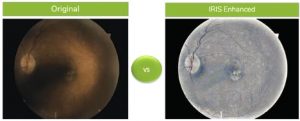
Historically, the gradability of traditional fundus images, captured using typical non-mydriatic cameras, has hovered around 80-85%. This is largely due to the orange coloring and dark, shaded areas that make it challenging for interpreting providers to determine an accurate diagnosis. IRIS image enhancement algorithms strip the fundus images of their coloring and illuminate the vasculature and pathology.
IRIS recently hosted a webinar with guest speakers from Austin Retina Associates who also serve as interpreting providers with the IRIS Reading Center. When asked to speak on the benefits of IRIS’s image enhancement feature, Dr. Jose Agustin Martinez shared “the enhanced image does a great job of uncovering the possibility of lipid underneath the dark shadow. This is why I love the IRIS platform unlike other platforms that I do read for, the IRIS platform really does make the photos more gradable.”
A high gradability rate is crucial because we have consistently found that roughly 1 in 10 diabetic patients who receive an IRIS retinal evaluation will have sight-threatening pathology that could result in vision loss if left untreated. Therefore, a higher gradability rate helps to ensure the patients with the sight-threatening disease are diagnosed and referred for treatment as quickly as possible. IRIS clients have an average gradability of 95%.
IRIS Chief Medical Officer, Ronald L. Gross, MD, explains “These images are being captured in a non-eyecare setting. This means the camera operators are not experienced ophthalmic technicians, these are people present in primary care offices. If you don’t capture an image that you can evaluate, That means the patient will need to go to an eye doctor for the exam.” To solve for getting the highest gradability, as a routine part of IRIS implementations with a new client, extensive onsite or virtual training is provided to camera operators, ensuring the best image capture techniques are mastered. Additionally, IRIS offers clients a vast library of refresher training videos that are accessible anytime post–go–live.
It’s clear that consistently high image gradability is a key factor in the success of a diabetic retinal exam program. That’s why IRIS created its proprietary enhanced image feature and why we will continue to find ways to increase accessibility to this exam.
To learn more about IRIS image enhancement, check out this video from our webinar with Austin Retina Associates.
Click here for more information on the IRIS Solution or contact us to schedule a demo!
SM053 Rev A
Get started with IRIS today.
Want to know if IRIS is right for you? Schedule a one-on-one consultation with our team. We’re here to help.